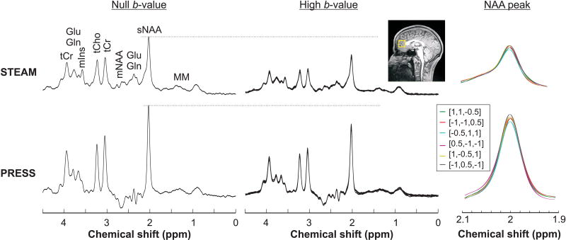
Figure 3.
DW spectra acquired from one subject using STEAM (TE/TM = 21.2/105 ms) and PRESS (TE = 54.2 ms) sequences. Spectra acquired at null b-value (left panel) and at high b-value in all six directions (middle panel) are shown. Excellent spectral quality, with no lipid contaminations and flat baseline were obtained in the prefrontal lobe VOI (yellow box on the T1-weighted image) with both techniques. The effects of cross-terms from the DW gradients are visible as the variation of the NAA amplitude at 2 ppm (right panel).

