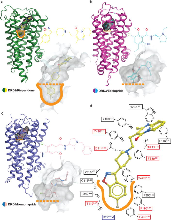
Figure 2. Comparison of the ligand binding pocket across the D2-like family receptors.

a, b, c, Surface representations of the ligand binding pockets of DRD2 (a), DRD3 (b) (PDB code 3PBL) and DRD4 (c) (PDB code 5WIU) are shown in transparent gray. d, A schematic representation of risperidone binding interactions at a 4.0 Å cut-off is shown. Hydrogen bonds are shown in grey dashed lines. Mutations of the amino acid in the red boxes reduces risperidone binding affinity by more than tenfold. The thermo-stabilizing mutation (I1223.40A) colored in blue. The outline of deeper hydrophobic pocket is colored as orange.
