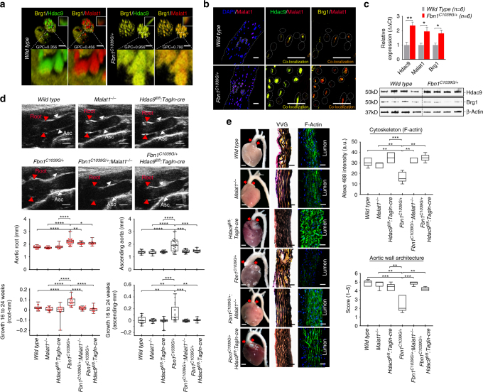
Fig. 6.
Genetic disruption of Malat1 or Hdac9 improves experimental aortic aneurysm. a Explanted VSMCs from Fbn1C1039G/+ mouse aortas demonstrate increased colocalization of Hdac9–Brg1 as well as Malat1–Brg1 when compared to wild-type cells. Global Pearson’s colocalization (GPC) of spatial overlap between Hdac9–Brg1 and Malat1–Brg1 is shown. Bar = 2.5 µM b Identification of Hdac9–Malat1–Brg1 complex in wild type and Fbn1C1039G/+ by confocal microscopy of ascending aortic samples. Bar = 40 µM c QPCR of Hdac9, Malat1, and Brg1 demonstrate upregulation in Fbn1C1039G/+ aortas, bar graphs are presented as mean with error bars (±S.D.) n = 6 animals, 6 months of age (d) Improved quantitative aortic dimensions in Fbn1C1039G/+:Malat1−/− and Fbn1C1039G/+:Hdac9fl/fl:Tagln-cre mice when compared to Fbn1C1039G/+ mice. Ultrasound of aortic root (Red) and ascending aortic (Black) quantification of wild-type (n = 18), Malat1−/− (n = 18), Hdac9fl/fl:Tagln-cre (n = 17), Fbn1C1039G/+ (n = 24), Fbn1C1039G/+:Malat1−/− (n = 19), and Fbn1C1039G/+:Hdac9fl/fl:Tagln-cre (n = 15; One-way ANOVA, *p < 0.05, **p < 0.01, ***p < 0.001). Aortic dimensions at 6 months and aortic growth between 16 and 24 weeks are shown. Representative images of parasternal long axis systolic ultrasound images of the aortic root of 6-month old mice. Red arrows denote aortic root, white arrows denote ascending aorta. Bar = 1 mm (e) Malat1 and Hdac9 deficiency rescues anatomic and cytoskeletal defects in Fbn1C1039G/+ aortas. Representative photomicrographs of six month old latex-injected wild-type, Malat1−/−, Hdac9fl/fl:Tagln-cre, Fbn1C1039G/+, Fbn1C1039G/+:Malat1−/−, and Fbn1C1039G/+:Hdac9fl/fl:Tagln-cre hearts and ascending aortas (left column, Bar = 1 mm, middle and right column, Bar = 40 µM). Red arrows indicate the ascending portion of the aorta. Aortas stained with Verhoeff-Van Gieson stain (center panels), and immunofluorescence of F-actin (right panels). The aortic adventitia faces left while the aortic lumen is oriented to the right. Quantification of aortic architecture and F-actin staining, (One-way ANOVA, *p < 0.05, **p < 0.01, ***p < 0.001). Full-length western blots presented in Supplementary Fig. 8

