Figure 4.
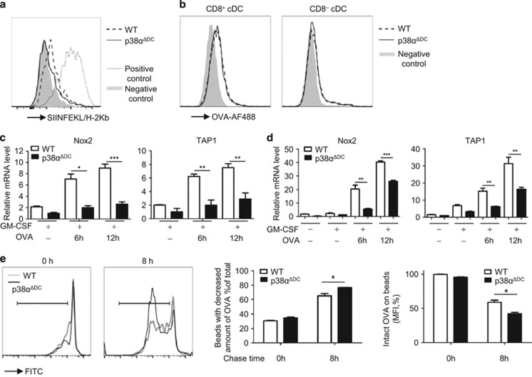
p38α had an important role in Ag processing. (a) Direct staining for the OVA peptide and MHC-I complex on the cell surface. The splenic CD8+ cDCs were incubated for 2 h with or without the OVA protein, washed, cultured for an additional 20 h, and then stained with SIINFEKL-H2Kb-biotin and streptavidin-PE. The dashed (WT) and solid (p38αΔDC) lines indicate staining with Ag pre-incubation; the gray shadow indicates background staining without Ag present during the pre-incubation (negative control) and the dotted line indicates staining with OVA257–264 pre-incubation (positive control). (b) Ag uptake into the cells. The splenic CD8+ or CD8− cDCs were incubated for 30 min at 37 °C with AF488-labeled OVA protein (5 μg/ml). The gray shadow indicates the negative control. The DCs were kept on ice for 10 min and then incubated with, OVA-AF488 on ice for 30 min. (c, d) Real time-PCR analysis of the of Nox2 and TAP1 mRNA levels in splenic CD8+ cDCs (c) and BM-derived CD24+ cDCs (d) from p38αΔDC mice and WT mice treated with GM-CSF (20 ng/ml) and OVA (100 μg/ml) as indicated. (e) Ag degradation was increased in the p38α-deficient CD24+ cDCs. The BM-derived CD24+ cDCs (1 × 10⁶) were cultured with OVA-coated latex beads (3 × 10⁶) for 30 min to allow phagocytosis, followed by removal of the free beads as described in the Methods. Then, the CD24+ cDCs were disrupted by lysis buffer immediately (0 h) or after another 8 h of culture (8 h). The recovered beads from cell lysis were stained with a FITC-conjugated anti-OVA antibody and the remaining OVA protein on beads was analyzed by flow cytometry. (a, b) The cDCs were sorted from five pairs of littermate mice. One of the two experiments with similar results is presented. The medium used was RPMI 1640 medium supplemented with 10% FBS, 1% P/S (penicillin and streptomycin) and GM-CSF (20 ng/ml). (c) The CD24+ cDCs were generated from BM cells from three pairs of littermate mice. (d) The CD8+ cDCs were sorted form 15 pairs of mice. (c, d) The data were representative of two independent experiments, each including triplicate samples. (e) The CD24+ cDCs were generated by BM cells from three pairs of littermate mice. The data were representative of two independent experiments, each including two repeats. The results were presented as the mean±s.e.m. *P<0.05; **P<0.01; ***P<0.001.

