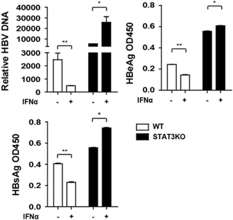
Figure 5.
HBV replication was increased in STAT3 knockout cells. HepG2 WT and STAT3KO cells were transfected with pHBV1.3. At 48 h, the cells were treated with IFNα or untreated, and after 24 h treatment, the supernatant was collected and HBV DNA, HBsAg and HBeAg in the supernatant were analyzed using Q-PCR or enzyme-linked immunosorbent assay. Data represent the means and s.d. from three independent experiments. Student’s t-test was performed. *P<0.05, **P<0.01.

