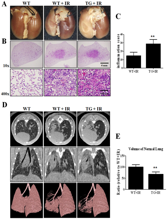
Figure 7.
Gross morphological, histopathological and micro-CT analysis in HSP27 transgenic mice. (A) Representative gross finding. Mice were sacrificed at two weeks after irradiation. Four-percent paraformaldehyde was instilled via trachea, and lungs were immersed in fixation solution and photographed after complete fixation. (B) Haematoxylin and eosin-stained lung sections. (C) Graphs show quantification of inflammation score. (D) Representative micro-CT images of lungs of irradiated and control mice. Horizontal (top row), trans-axial (middle row), and 3D micro-CT (bottom row) images acquired at two weeks after irradiation. (E) Quantification of volume of normal left lung was presented as mean ± standard error. **P < 0.01 versus BL6 IR.

