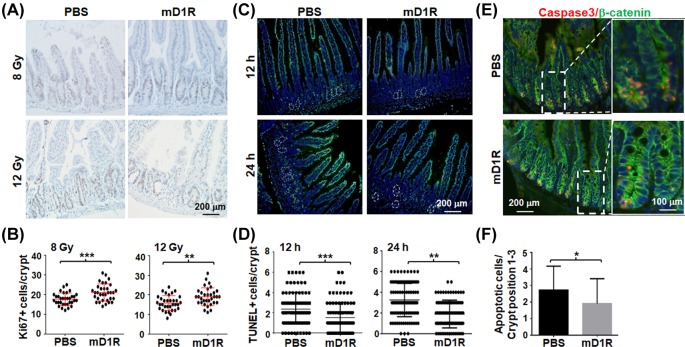
Figure 2. mD1R enhanced the proliferation and reduced the apoptosis of intestinal cryptic cells after irradiation in mice.
(A and B) Mice were irradiated by TBI with 8 or 12 Gy of γ-ray and injected daily i.p. with PBS or 100 μg of mD1R from the first day after the irradiation. On the third day after irradiation, the intestine of the mice were collected, and stained by immunohistochemistry with anti-Ki67 (A). The number of Ki67+ cells per crypt was counted and compared between the two groups (B). (C and D) Mice were irradiated by TBI with 10 Gy of γ-ray and injected i.p. with PBS or 100 μg of mD1R immediately after the irradiation. The intestine samples of the mice were collected on 12 and 24 h after the irradiation and stained with TUNEL. Nuclei were counter-stained with Hoechst33258 (C). The number of TUNEL+ cells per crypt was counted and compared between the two groups (D). (E and F) Samples in (C) were stained by immunofluorescence with anti-Caspase 3 and anti-β-catenin. Nuclei were counter-stained with Hoechst33258 (E). The number of apoptotic cells per crypt was counted and compared (F); bars = mean ± SD (n=5); *P<0.05, **P<0.01, ***P<0.001.

