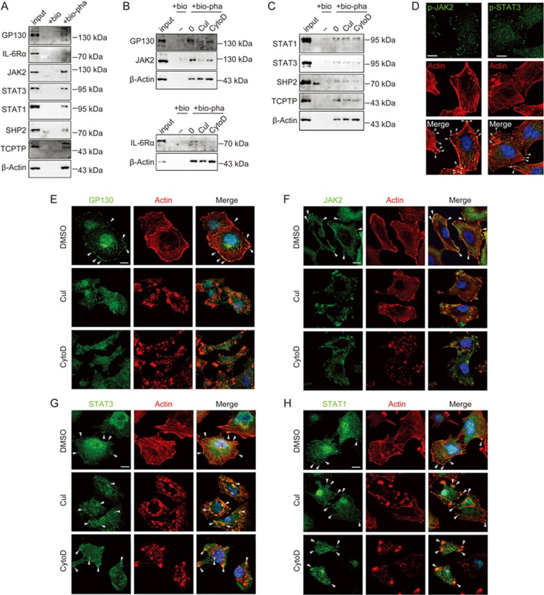
Figure 5.
STAT3 and STAT1 signaling proteins were physically associated with actin filaments. (A–C) A549 cells were lysed, and actin filaments were pulled-down using biotinylated-phalloidin, followed by elution with 10 mmol/L biotin. For drug treatments in B and C, A549 cells were treated with DMSO, 0.5 μmol/L CuI or 1 μmol/L CytoD for 2 h. input, 3% of the total cell lysates compared with 100% of the elution; bio, biotin; bio-pha, biotinylated-phalloidin. (D–H) Confocal micrographs of A549 cells staining with rhodamine-phalloidin (for actin, red), DAPI (for nucleus, blue), and antibodies against the indicated proteins (green). Arrowheads indicated colocalization of STATs signaling proteins with actin. For drug treatment, A549 cells were treated with DMSO, 0.5 μmol/L CuI or 1 μmol/L CytoD for 2 h prior to immunochemical staining. Scale bars, 10 μm.

