Figure 1.
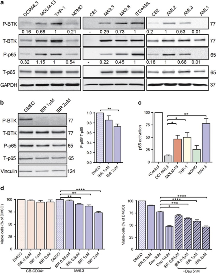
BTK and p65 are activated in MLL-rearranged cells and pharmacological inhibition of BTK induced cell death. (a) AML cell lines OCI AML3, MOLM-13, THP-1, NOMO (left), cord blood CD34+ cells (CB1), MA9 clones (MA9.3 and MA9.6), FLT3-ITD+AML (ITD+AML) patient leukemic blasts (middle) and CB2, MLL+AML patient leukemic blasts (AML1, 2 and 3; right) were lysed and immunoblotted for phospho-p65 (pS536) and -BTK (pY223) and total (T) proteins. GAPDH is internal loading control. ITD+AML cell lysates served as a positive control for the detection of phosphorylated p65 and BTK. Densitometric analysis of phosphorylated BTK and p65 relative to total proteins is indicated under the blots. (b) MA9.3 cells were serum-starved for 3 h and subsequently treated with 1 and 2 μm ibrutinib (IBR) for additional 2 h. Whole cell lysates were analyzed for the activation of p65 and BTK proteins. Vinculin is internal loading control (left). Densitometric analysis of P-p65 relative to T-p65 from three independent experiments is shown (right). (c) OCI AML3, MOLM-13, THP-1, NOMO and MA9.3 cells were lysed and transcriptionally active p65 is detected via NFκB activation assay following the manufacturer’s instructions. Nuclear extracts from Jurkat cells stimulated with calcium ionophore and tissue plasminogen activator served as positive (+) control. p65 activation from three independent experiments (in duplicates) is plotted. (d) Control CB-CD34+ cells and MA9.3 cells were treated with DMSO or different doses of ibrutinib (IBR) (left) or daunorubicin (DAU) alone or in combination (right) as indicated for 48 h and assayed for cell viability. Percentage of viable cells (Annexin V and sytox blue negative) from four independent experiments is shown. Error bars indicate s.e.m. *P<0.05, **P<0.01, ***P<0.001 and ****P<0.0001.

