Figure 2.
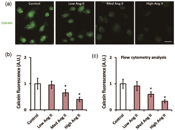
Angiotensin II (Ang II) facilitates mitochondrial permeability transition pore (mPTP) opening in CATH.a cells. CATH.a cells were treated with different concentration of Ang II (5, 50, and 500 nM) for 24 h, and (a) and (b) illustrate the CoCl2-calcein fluorescence quenching assay which was employed to evaluate mPTP opening. Note that the calcein fluorescence was decreased after incubation with Ang II. (c) The calcein fluorescence was further evaluated by flow cytometric analysis. All figures are representative of three independent experiments, performed in triplicate. Data were analyzed by one-way analysis of variance (ANOVA) followed by Tukey’s post-hoc test. Columns represent mean±standard deviation (SD). *p<0.05 versus control group.

