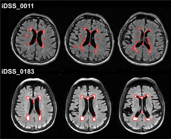
Fig. 1.

Two iDSS subjects with similar total WMH volume (iDSS_0011: 18.3 ml and iDSS_0183: 18.6 ml), but different confluency sum score (COSU) of the WMHs (21.9 and 8.8, respectively). The performance in the trail making test Awas more strongly impaired in the subject with the larger COSU (z-score −2.4 and −1.0, respectively). The red contour represents the automatically segmented white matter hyperintensities overlaid to the individual FLAIR image in native space. Three consecutive slices at the level of the inner ventricles are displayed for each subject
