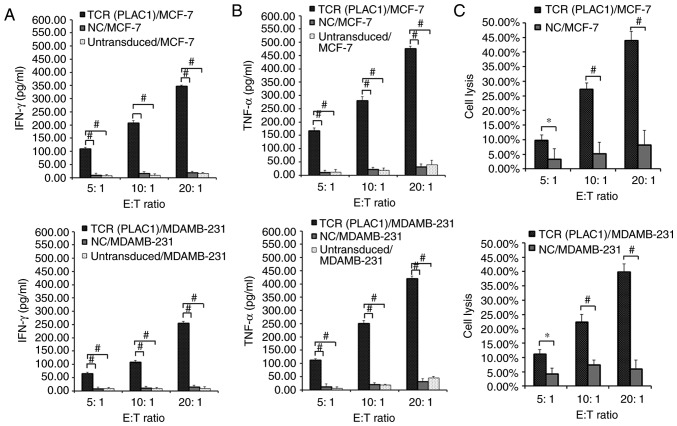
Figure 3.
Evaluation of the function of PLAC1 TCR-engineered CD8+ T cells. Human leukocyte antigen-A2-restricted and PLAC1-specific TCR-, NC-transduced or untransduced CD8+ T cells were co-cultured for 4 h with 1×10⁴ MCF-7 and MDAMB-231 cells respectively at a range of E:T ratios (5:1, 10:1 and 20:1). Concentration of (A) IFN-γ and (B) TNF-α secreted into the culture medium were measured using an enzyme-linked immunosorbent assay. (C) TCR- or NC-transduced CD8+ T cells were co-cultured for 4 h with 1×10⁴ MCF-7 and MDAMB-231 cells. Cytolysis was determined using a lactate dehydrogenase activity assay. Subsequent to background subtraction, the percentage lysis was calculated by 100% × [(experimental release-effector spontaneous release-target spontaneous release)/(target maximum release-target spontaneous release)]. *P<0.05 and #P<0.01 with comparisons shown by lines. PLAC1, placenta-specific 1; TCR, T cell receptor; CD8+ T cell, cytotoxic T cell; NC, negative control; E:T, effector cell to target cell; IFN-γ, interferon γ; TNF-α, tumor necrosis factor α.

