
Figure 1.
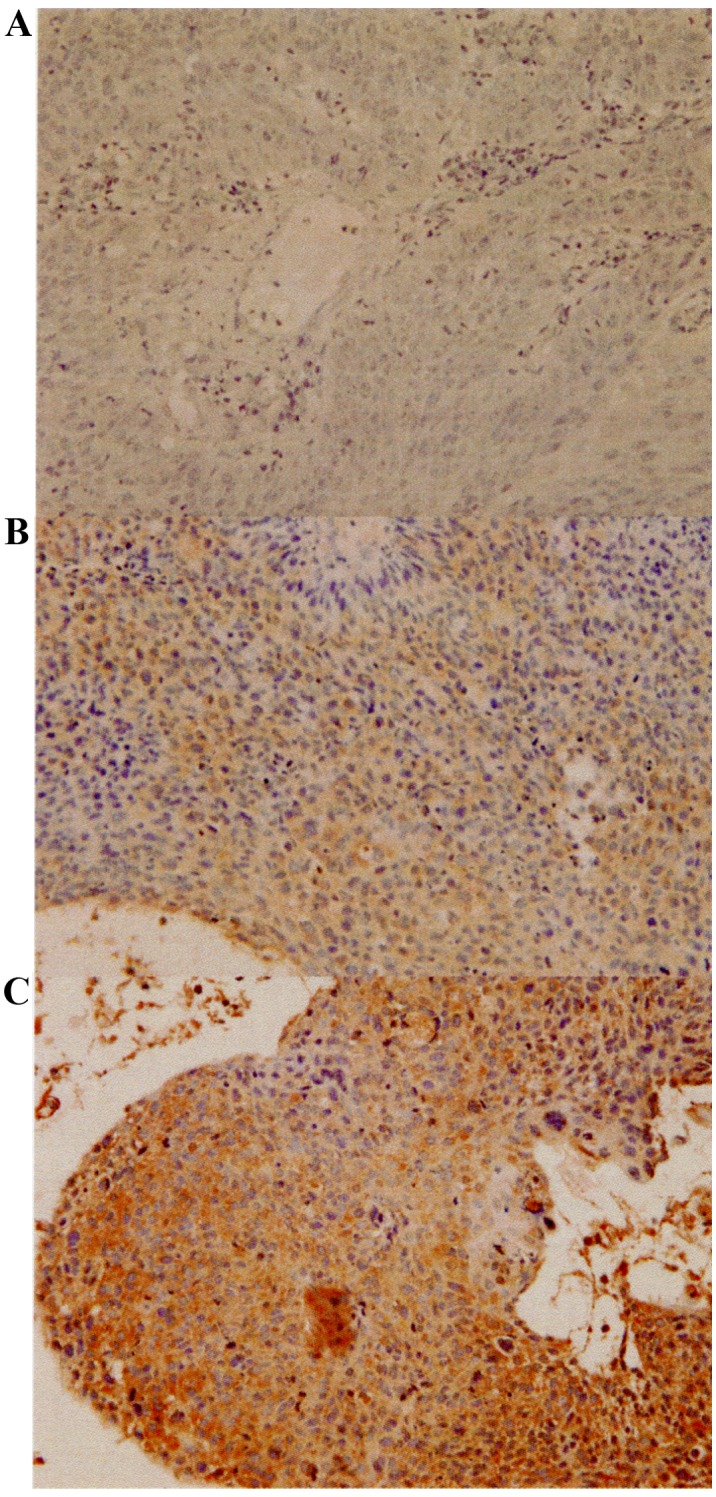
Immunohistochemical staining of XPA in locally advanced cervical cancer. (A) Negative control staining performed without primary antibody. (B and C) Representative sections stained with a primary antibody against XPA showing scores of 6 (B) and 12 (C). Sections were counterstained with hematoxylin. Magnification, ×400. XPA, xeroderma pigmentosum complementation group A.