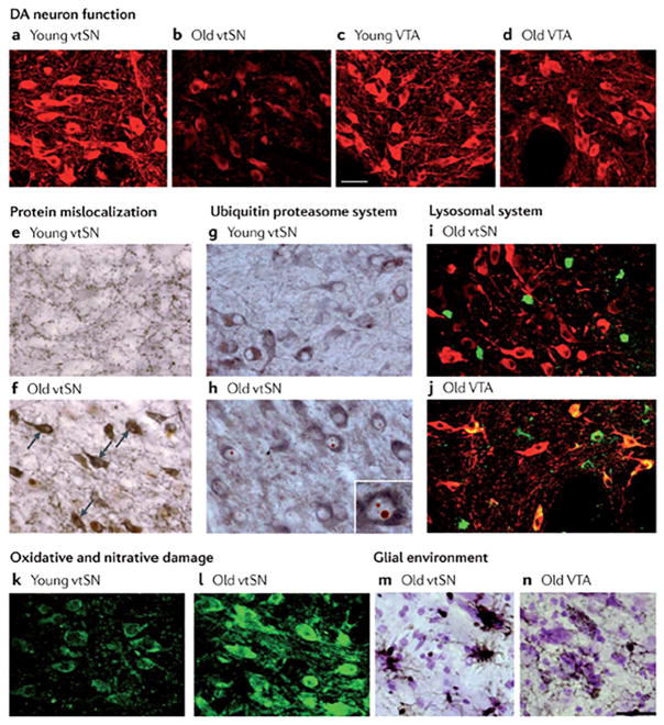
FIG. 1.
The pattern of ageing-related changes in markers of cellular mechanisms. With advancing age, DA neurons in the ventral tier of the substantia nigra (vtSN)—the population that is most vulnerable to degeneration in PD—show changes with aging. a–d: Age-related decline in tyrosine hydroxylase staining (shown in red) in vtSN neurons, but not in vental tegmental area (VTA) neurons. e,f: Accumulation of cytoplasmic α-synuclein (shown in brown). Tyrosine hydroxylase staining is shown in gray. Arrows show examples of cytoplasmic α-synuclein in aged vtSN. g,h: Increased numbers of Marinesco bodies, characterized by cytoplasmic inclusions of ubiquitin (shown in red). Tyrosine hydroxylase staining is shown in gray. The inset (in part h) is a higher magnification view of a tyrosine hydroxylase immunoreactive neuron of the vtSN exhibiting multiple Marinesco bodies. i,j: No accumulation of lipofuscin (shown in green). Tyrosine hydroxylase staining is shown in red, colocalization of lipofuscin and tyrosine hydroxylase is shown in yellow. Note that virtually all lipofuscin staining in the vtSN is not in dopamine neurons, whereas colocalization is apparent in aged VTA neurons. k,l: Accumulation of 3-nitrotyrosine (shown in green). m,n: Greater microglial reactivity in aged vtSN neurons than in aged VTA neurons, shown by greater staining for human leukocyte antigen (HLA) class II histocompatibility antigen, antigen D related (DR) α-chain (HLA-DRA; a marker for microglia), shown in brown. UPS, ubiquitin–proteasome system. From Ref. 5 with permission.

