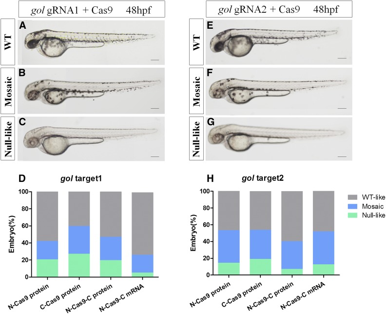
Figure 6.
Mutagenetic phenotype of various nuclear localization signal (NLS)-fused Cas9 proteins and N-Cas9-C mRNA for two sites of the gol gene. (A–C) and (E–G) show lateral views of wild-type (WT) (A and E), gol-target 1 (B and C), and gol-target 2 (F and G) at 48 hpf (hr postfertilization). The phenotypes observed were WT (A and E), mosaic retinal pigmented epithelium (RPE) (B and F), and unpigmented RPE (C and G). (D and H) Proportions of embryos with each phenotype. Scale bars: 0.2 mm.

