Figure 2.
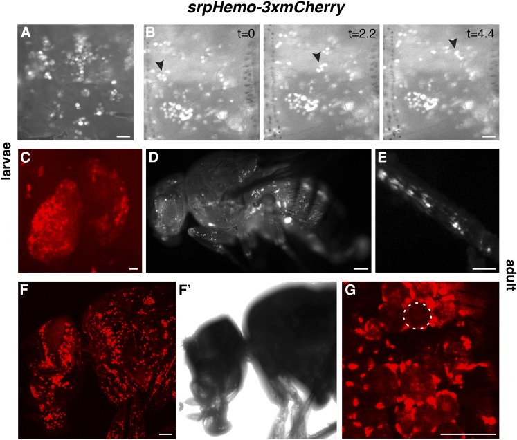
Direct fusion lines allow live imaging of plasmatocytes in larvae and adults. (A) Live image of plasmatocytes sitting on the body wall of a srpHemo-3xmCherry larva, viewed through the cuticle with a stereomicroscope. (B) Time-lapse imaging of plasmatocytes in a srpHemo-3xmCherry larva filmed through a stereomicroscope. Three successive time points separated by 2.2 sec each are shown. Arrowhead indicates a group of cells that float in the hemolymph while most other cells remain attached to the body wall. (C) Confocal image of labeling by srpHemo-3xmCherry third-instar larval lymph gland. (D) Live image of plasmatocytes in a srpHemo-3xmCherry adult and in a close-up of (E) the leg viewed through a stereomicroscope. (F–G) Live image of a srpHemo-3xmCherry adult viewed with a confocal microscope. (F) 3D projection of plasmatocytes in the head, proboscis, and thorax. (F’) transmitted light view of the adult fly imaged in (F). (G) Single confocal slice showing plasmatocytes encircling adult fat body cells (one indicated with white circle). Scale bars correspond to 500 μM in (A–C), 250 μM in (D and E), and 100 μm in (F–G).

