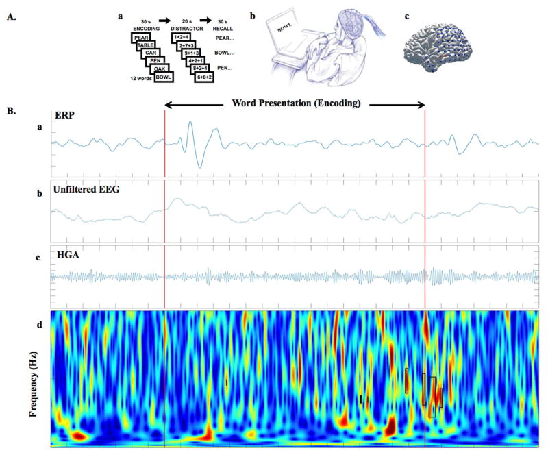
Figure 2. A) Cognitive task paradigm for studying physiological nHFOs.
Patients with epilepsy who are implanted with subdural and depth electrodes for seizure monitoring as part of the clinical evaluation for drug-resistant epilepsy provide a unique opportunity for neuroscientists to perform cognitive tasks and record the electrophysiological signals directly from the human brain. Areas of active research include investigation of the neural correlates of cognition. These cognitive tasks may help to improve the discrimination of nHFOs from pHFOs. In our study, we used iEEG recorded during a verbal memory task in 11 epilepsy patients to analyze gamma frequency events within and outside the seizure generating brain regions. A. a) Diagram of free recall verbal memory task, A. b) Epileptic patients participating in the study during second phase monitoring doing verbal memory tasks on a laptop, A. c) brain surface of an example patient with implanted grid and strip electrodes the iEEG signal is recording from (unpublished data). B) From top to bottom, B. a) shows the trial-averaged ERP signal for a whole session (300 words presented), plots below are raw data from individual example trial during same session, that subject subsequently recalled the presented word. From top to bottom, the unfiltered EEG (fig 2. B. b), the band-pass filtered signal for high gamma activity (HGA) (fig 2. B. c), and the continuous time frequency image with detected HGAs highlighted with black rectangles (fig 2. B. d). Red lines are stimulus (word presentation) onset and offset (F Khadjevand et al., unpublished).

