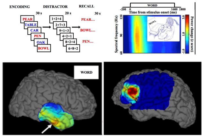
Figure 3. Brain mapping using physiologic task-induced HFOs and pathological HFOs Top, left).
Word lists are presented one-by-one for encoding & subsequent recall. Presentation of words induces high gamma activities (60–120 Hz) in specific brain areas. In this figure subsequently recalled words printed in red and forgotten words presented in blue. Top, right) Spectrogram of local field power during encoding epochs aligned to the presented word. Bottom) Left panel, brain surface maps of high physiological gamma power interpolated over 4×6 temporal cortex grid (White arrow focus of high activation) and in right panel the pathological HFOs over the epileptic region of the brain.

