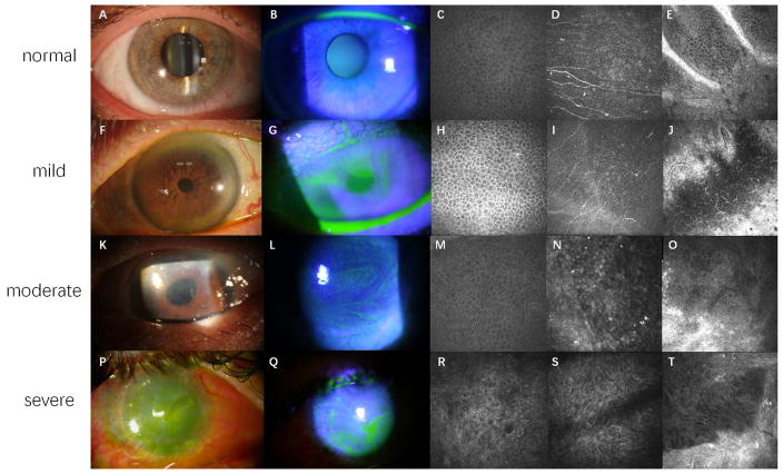
Figure 1.
Clinical presentations and confocal images of LSCD at different stages.
Figure 1A, 1F, 1K and 1P are slit lamp photos under white light. Figure 1B, 1G, 1L and 1Q are slit lamp photos under cobalt blue light. Figure 1C, 1H, 1M and 1R are confocal images of wing cells at central cornea. Figure 1D, 1I, 1N and 1S are confocal images of epithelial basal cells at central cornea. Figure 1E, 1J, 1O and 1T are confocal images of limbal epithelium and the palisades of Vogt.
Healthy corneal epithelium is transparent (A) without fluorescein staining (B). Normal wing cells (C) have a dark cytoplasm, well-defined bright borders, and no visible nuclei. Basal epithelial cells (D), which are characterized with dark cytoplasm, fairly defined cell borders and no visible nuclei, are clearly seen in normal eyes. The subbasal nerve plexus is also visible in this layer. In healthy eyes, the palisades of Vogt may be detected at the limbal area and appear as hyper-reflective, double-contour, linear structures (E). Late fluorescein staining can be detected at mild stage of LSCD, with a clear line of demarcation may be visible between the corneal and conjunctival epithelial cells in sectoral LSCD (F and G). Corneal epithelial cells in mild LSCD have less distinct borders and prominent nuclei (H and I). The density of subbasal nerve plexus decrease dramatically, along with the infiltration of dendritic cells (I). The structure of palisades of Vogt is altered even in mild LSCD. A large number of inflammatory and dendritic cells are sometimes present with blood vessels (J). Epithelial opacity (K) and persistent late fluorescein staining in a vortex pattern (L) are the signs of a more advanced stage. The morphological abnormalities of wing cells (M) and basal cells (N) at central cornea continue to progress. The degradation of subbusal nerve plexus deteriorates further (N). The palisades of Vogt are absent at limbal area, along with enlargement of limbal epithelial cells (O). Epithelial and stromal opacity, neovascularization (P), and persistent epithelial defect (Q) on the corneal surface may be present in severe or total LSCD. Metaplastic epithelial cells are visible in eyes with severe LSCD (R and S). The structures of limbus and palisades of Vogt are totally damaged (T).

