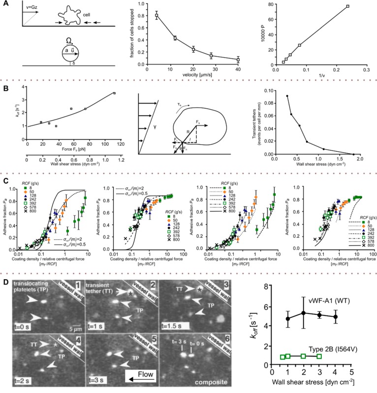
Figure 16.
Shear effects on receptor–ligand binding kinetics. (A) Studies of the kinetics of bond formation and dissociation of activated neutrophils and endothelial cells on the surface on a flow chamber. A minimal shear drag force was exerted that was lower than the strength of a single molecular bond. The graph shows the observed relation of the probability of neutrophile adhesion to the bilayer and the flow velocity. x Axis: (flow velocity)−1, y axis: probability of adhesion. (B) In a flow chamber, binding and unbinding events of neutrophils flowing over a bilayer containing P-selectin tethers were monitored to understand the biophysics of cell rolling. The koff values shown in the graph were calculated by comparing the binding rates in shear stressed and nonstressed systems. x Axis: shear stress vs off-rate; y axis: koff (s–1). (C) Comparison of the abilities of the two models to account for the data. Predictions (curves) of model II (a and b) and model I (c and d) for the unsaturated (a and c) and saturated (b and d) bindings. (D) Dependence of koff on the shear stress for WT and mutant substrates. On the basis of Goldman’s equations, the force acting on a platelet in shear flow was calculated. (A) Adapted with permission from ref (207). Copyright 1993 Elsevier. (B) Adapted with permission from ref (208). Copyright 1995 MacMillan Publisher (C) Adapted with permission from ref (209). Copyright 1998 Elsevier. (D) Adapted with permission from ref (210). Copyright 2002 Elsevier.

