. 2018 Mar 9;7:e33033. doi: 10.7554/eLife.33033

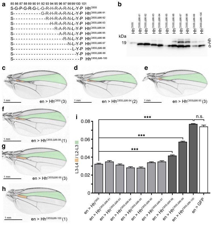
Figure 4. N-terminal truncation of palmitoylation-deficient HhC85S rescues wing formation.

(a) All truncated proteins also lacked the N-terminal cysteine, preventing Hh palmitoylation (Hardy and Resh, 2012). Residues #93–97: CW motif. (b) All proteins were expressed and secreted from S2 cells, as determined by immunoblotting. (c–h) En-regulated overexpression of HhC85S and N-terminally truncated proteins (HhC85S;Δ). Unaffected wing development despite en-regulated expression of unpalmitoylated HhC85S;Δ86-100 (h). (i) Quantification of wings shown in c-h. En-regulated GFP and HhC85S expressions served as positive and negative controls, respectively. Pooled analysis of three transgenic fly lines, each derived from an independent injection. en >HhC85S: 0.032 ± 0.001, en >HhC85S;Δ86-91: 0.035 ± 0.001 (p=0.1375), en >HhC85S;Δ86-93: 0.031 ± 0.001 (p=0.5458), en >HhC85S;Δ86-94: 0.028 ± 0.001 (p=0.0134), en >HhC85S;Δ86-95: 0.028 ± 0.001 (p=0.001), en >HhC85S;Δ86-96: 0.034 ± 0.001 (p=0.25), en >HhC85S;Δ86-97: 0.035 ± 0.001 (p=0.117), en >HhC85S;Δ86-98: 0.041 ± 0.001 (p<0.0001), en >HhC85S;Δ86-99: 0.057 ± 0.001 (p<0.0001); en >HhC85S;Δ86-100: 0.076 ± 0.0007 (p=0.0001), en >GFP: 0.074 ± 0.002. ***p≤0.001, n.s. (not significant): p>0.05, n = 60 (n = 20 per line), all flies developed at 25°C.

Figure 4.

Figure 4—figure supplement 1. Modeled linear Drosophila Hh clusters, using pdb 2IBG and pdb 3M1N as templates.
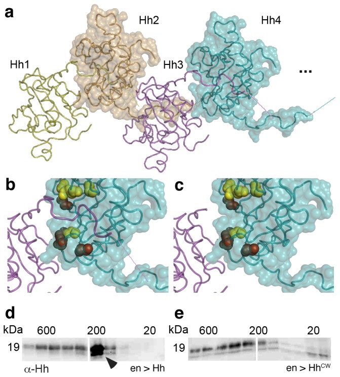
Figure 4—figure supplement 1.

(a) Predicted intermolecular Drosophila Hh interactions, resulting in linear zigzag chains. The molecular surface of every other Hh molecule is shown to demonstrate intermolecular interactions between N-terminal palmitoylated peptides and the adjacent molecules in the chain. Modeled N-terminal palmitate is shown as a line (pointing to the right). (b) Close-up of Hh N-terminal interactions with the predicted Ptc binding site of the adjacent molecule in the cluster. Yellow spheres denote Drosophila Hh residues corresponding to Shh residues that interact with Ptc (Drosophila Hh H193, H194, H200, H240) (Bosanac et al., 2009). Red spheres denote residues corresponding to Shh amino acids bound by the Shh inhibitory antibody 5E1 (K105, R147, R213, R238, R239 in Drosophila Hh) (Maun et al., 2010). (c) Based on our model, N-terminal Hh truncation makes these sites accessible in the cluster. (d,e) Top: Gel filtration analysis of transgenic L3 fly larvae expressing Hh or a HhCW variant with a mutated Hh cleavage site under en-control in vivo. Note the presence of a truncated Hh fraction (representing the situation in c) in larvae expressing ectopic wild-type Hh (arrowhead). Top bands in d,e represent the situation shown in b, bottom bands the situation shown in c.
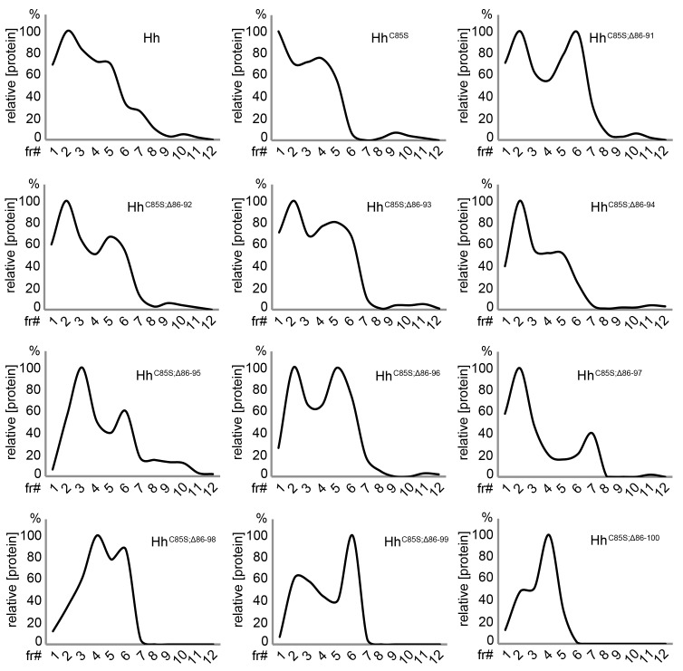
Figure 4—figure supplement 2. Unimpaired multimerization of N-terminally truncated Hh variants.

Figure 4—figure supplement 2.

Gel filtration analysis of Hh, HhC85S, and N-terminally truncated variants (HhC85S;Δ86-91 - HhC85S;Δ86-100) expressed in Drosophila S2 cells under actin-control. Soluble Hhs were detected in the form of ‘large’ (>600 kDa, fractions 1–2) and ‘smaller’ (100 kDa-600 kDa, fractions 3–6) clusters. Elution profiles are expressed relative to the highest protein amounts in a given fraction, which was set to 100%.
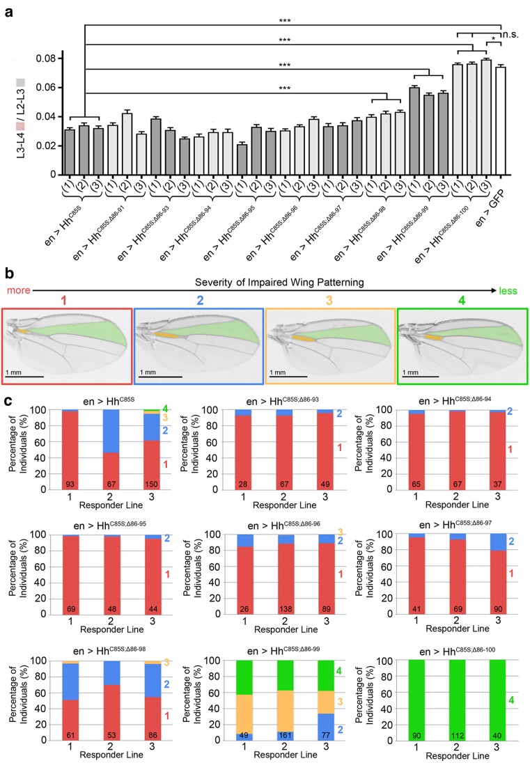
Figure 4—figure supplement 3. Graded variable wing defects as a consequence of full-length and N-terminally truncated HhC85S expression in the wing disc.

Figure 4—figure supplement 3.

(a) Analysis of three transgenic fly lines, each derived from an independent injection of cDNA encoding the same unpalmitoylated N-truncated construct. En-regulated GFP and HhC85S expressions served as positive and negative controls, respectively. en>HhC85S (1-3): 0.032 ± 0.001, en>HhC85S;Δ86-91 (1-3): 0.035 ± 0.001 (p=0.1375), en>HhC85S;Δ86-93 (1-3): 0.031 ± 0.001 (p=0.5458), en>HhC85S;Δ86-94 (1-3): 0.028 ± 0.001 (p=0.0134), en>HhC85S;Δ86-95 (1-3): 0.028 ± 0.001 (p=0.0085), en>HhC85S;Δ86-96 (1-3): 0.034 ± 0.001 (p=0.2503), en>HhC85S;Δ86-97 (1-3): 0.035 ± 0.001 (p=0.1165), en>HhC85S;Δ86-98 (1-3): 0.041 ± 0.001 (p<0.0001), en>HhC85S;Δ86-99 (1-3): 0.057 ± 0.001 (p<0.0001); en>HhC85S;Δ86-100 (1): 0.076 ± 0.001 (p=0.4058), en>HhC85S;Δ86-100 (2): 0.076 ± 0.001 (p=0.3330), en >GFP: 0.074 ± 0.002. ***p≤0.001, **p≤0.01, *p≤0.05, n.s. (not significant): p>0.05, n = 20 (per line), all flies developed at 25°C. (b) Observed wing phenotypes were classified into four distinct grades, ranging from 1 (strongly affected wings, red) to 4 (wild-type wings, green). (c) Variations in wing formation are expressed as relative percentages of observed phenotypes, using the scheme shown in a. Three transgenic fly lines derived from independent injections of each construct were analyzed (1-3); the number of analyzed wings is shown in the bars. Note the phenotypic variations between transgenic lines of the same construct, which we explain as stochastic differences in the relative amounts and distributions of unpalmitoylated transgenic proteins in wild-type Hh clusters. A significant fraction of wings obtained from en>HhC85S;Δ86-99 flies were normal, and all wings from en>HhC85S;Δ86-100 flies were indistinguishable from wild-type wings. This demonstrates that dominant-negative HhC85S protein activities are fully reversed by the additional removal of N-terminal inhibitory peptides.
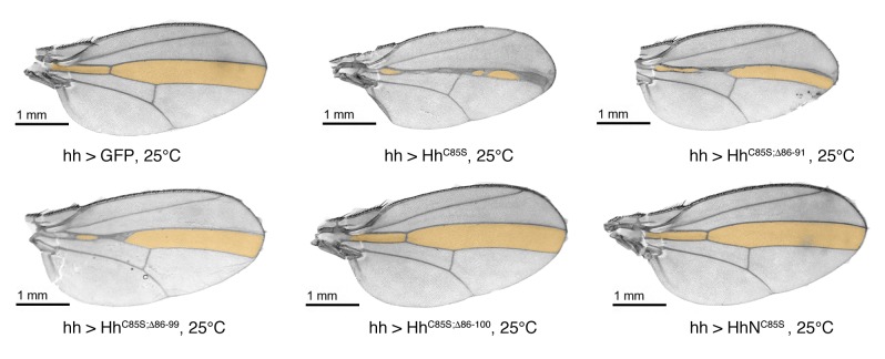
Figure 4—figure supplement 4. Confirmation that N-truncated HhC85S and HhNC85S do not repress the formation of Hh-dependent wing structures, using the driver line hh-Gal4.

Figure 4—figure supplement 4.

Confirmation of wing phenotypes obtained from en-Gal4-driven transgene expression by the alternative posterior driver line hh-Gal4. In contrast to multimeric, non-palmitoylated HhC85S, hh-Gal4 controlled expression of N-terminally truncated, palmitoylation-deficient HhC85S;Δ86-100 and unlipidated monomeric HhNC85S do not impair the Hh-regulated formation of the L3-L4 intervein region (orange). A hh >GFP control wing is also shown. Anterior is up, proximal is left.
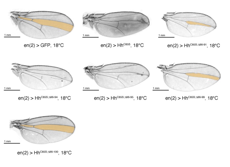
Figure 4—figure supplement 5. Confirmation that N-truncated HhC85S does not repress the formation of Hh-dependent wing structures, using the driver line en(2)-Gal4.

Figure 4—figure supplement 5.

Confirmation of wing phenotypes using the alternative posterior driver line en(2)-Gal4. As previously shown for en-Gal4 and hh-Gal4 driven transgene expression, non-palmitoylated HhC85S, but not N-terminally truncated, palmitoylation-deficient HhC85S;Δ86-100 reduce the L3-L4 intervein region (orange). An en(2) >GFP control wing is shown. Anterior is up, proximal is left.
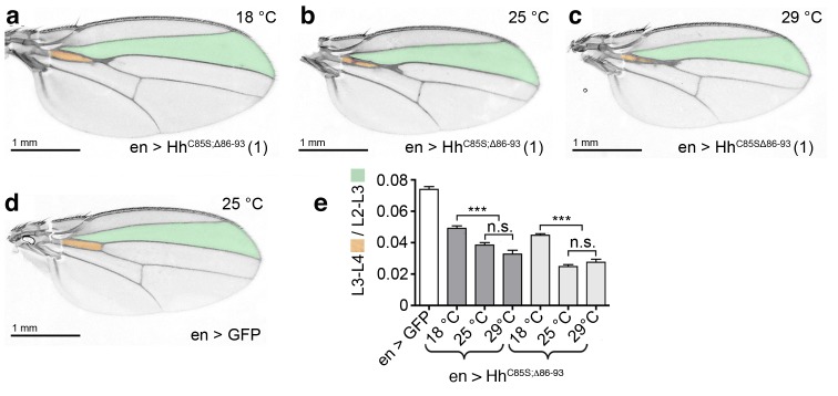
Figure 4—figure supplement 6. Temperature-dependent dominant-negative HhC85S;Δ86-93 function in the posterior Drosophila wing disc compartment.

Figure 4—figure supplement 6.

(a–c) en-driven overexpression of HhC85S;Δ86-93 in a wild-type background at 18°C, 25°C, and 29°C. Adult Drosophila wings are shown. (d) GFP served as control (wild-type wing). At 25°C and at 29°C, the L3-L4 intervein space of en>HhC85S;Δ86-93 wings narrows if compared with wings developing at 18°C. In addition, wings get smaller. (e) Quantification of wild-type Hh patterning activity. Two transgenic lines were analyzed (brackets). en >GFP: 0.074 ± 0.002, en>HhC85S;Δ86-93 (line 1) at 18°C: 0.049 ± 0.002, at 25°C: 0.038 ± 0.002, and at 29°C: 0.033 ± 0.002. Differences between 25°C and 29°C were insignificant (p=0.06); differences between 18°C and 25°C (p<0.0001) and 29°C (p<0.0001) were significant. en>HhC85S;Δ86-93 (line 3) at 18°C: 0.045 ± 0.001, at 25°C: 0.025 ± 0.001, and at 29°C: 0.028 ± 0.002. Differences between 25°C and 29°C were insignificant (p=0.2450); differences between 18°C and 25°C (p<0.0001) and 29°C (p<0.0001) were again significant. ***p≤0.0001; n.s. (not significant): p>0.05, n = 20.