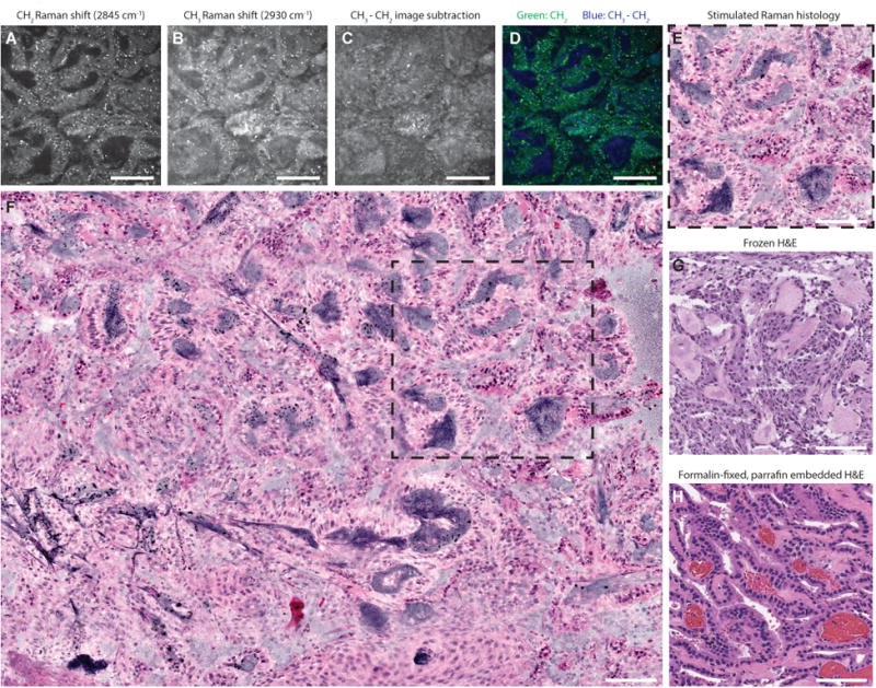
Figure 1. Label-free stimulated Raman histology (SRH) of fresh brain tumor tissue.

A choroid plexus papilloma, WHO grade I, imaged at 2845 cm-1 (A) and 2930 cm-1 (B) Raman shift wave numbers with 400 × 400-μm fields of view at a rate of 2 seconds per frame. To highlight nuclear contrast, 2930 cm-1 image is substracted from the 2845 cm-1 image in a single post-processing (C). Two-channel blue-green image (D) is generated by assigning blue gradient to the 2930-2845 cm-1 pixel intensity and green to the 2845 cm-1 pixel intensity. Our hematoxylin and eosin (H&E) color lookup table is applied to produce SRH (E) to emulate standard H&E staining of frozen (G) and formalin-fixed, paraffin-embedded (H) sections. SRH mosaics (F) are created by automated stitching of individual SRH tiles (dashed square). Scale bars are 100 μm.
