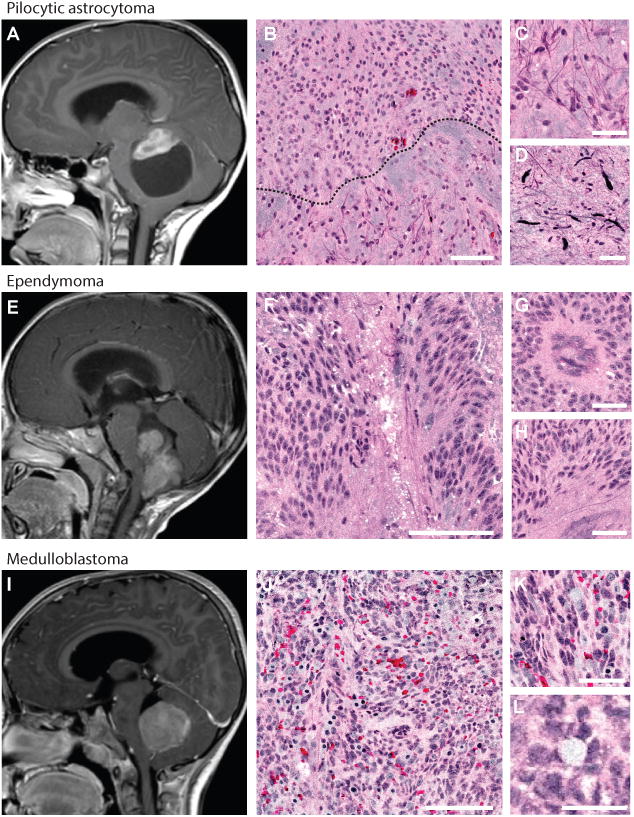
Figure 3. SRH identifies pediatric surgical lesions of the posterior fossa.

Magnetic resonance images (midsagittal T1-weighted post-gadolinium) of the three most common surgical lesions of posterior fossa are shown: pilocytic astrocytoma (A), ependymoma (E), and medulloblastoma (I). Pilocytic astrocytoma SRH shows biphasic pattern (black dashed line, B) with protein-rich pilocytic processes (C) and Rosenthal fibers (D). Ependymomas demonstrate rosette formation (F) and pseudorosette formation shown in cross-section (G) and longitudinal section (H). Medulloblastomas are densely hypercellular on low- (J) and high-magnification (K). Homer-Wright rosette formation (L) is visualized throughout the SRH mosaic. Scale bars are 100 μm in large tiles, 50 μm in small tiles.
