
Figure 4. SRH preserves cytologic and histoarchitectural features of pediatric brain tumors.
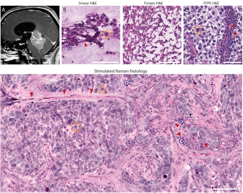
(A) Preoperative midsagittal T1-weighted post-gadolinium magnetic resonance image of posterior fossa germinoma. (B) Smear preparation shows the large germ cells with abundant foamy glycogen-rich cytoplasm (yellow circle), admixed with reactive small lymphocyte (blue circle) adjacent to blood vessels (red arrows). (C) Frozen sectioning causes freezing artifact that disrupts essential cytologic features of germinoma, severely limiting interpretation. (D) Formalin-fixed, paraffin-embedded (FFPE) H&E section shows large tumor cells with prominent nucleoli and mature lymphocytes adjacent to blood vessels (red arrows). (E) Similar to FFPE image, key diagnostic features are shown in SRH with preserved specimen cytology and histoarchitecture, allowing for unhindered interpretation and accurate histopathologic diagnosis.