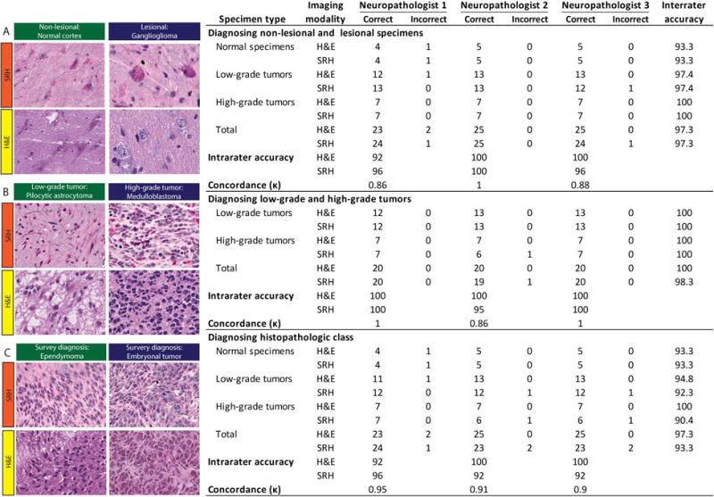
Figure 5. Evaluation of SRH via simulated intraoperative pathology consultation.

Results from web-based survey shown in the table. SRH and standard H&E images from 25 patients were presented to three neuropathologists for evaluation. Free-text responses were evaluated on three levels: 1) Normal versus lesional (A), 2) low-grade versus high-grade (B), and histopathologic diagnosis (C). Examples of images included in the survey are shown with corresponding SRH and H&E images above: normal cortex; ganglioglioma, WHO grade I; pilocytic astrocytoma, WHO grade I; medulloblastoma (MB), WHO grade IV; ependymoma, WHO grade II; and embryonal tumor other than MB, WHO grade IV.
