
Fig. 2. Structural characterization of nanowood.
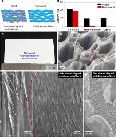
(A) Schematics of the aligned cellulose nanofibrils in the nanowood before and after which the intermixed amorphous lignin and hemicellulose have been removed. (B) Concentration of lignin, hemicellulose, and cellulose in the natural wood and nanowood. (C) Photograph of a nanowood specimen that exhibits pure bight color and an aligned texture. (D) Nanowood exhibits a large porosity, a hierarchical structural alignment of fibril aggregates, and a maintained alignment of the fibril aggregates. (E) Side-view SEM image of the microsized porous and aligned channels inside the nanowood. (F) SEM image of the porous channel walls that composed of aligned nanofibrils. (G) Top-view SEM image of the nanowood channels with separated nanofibrils ends.