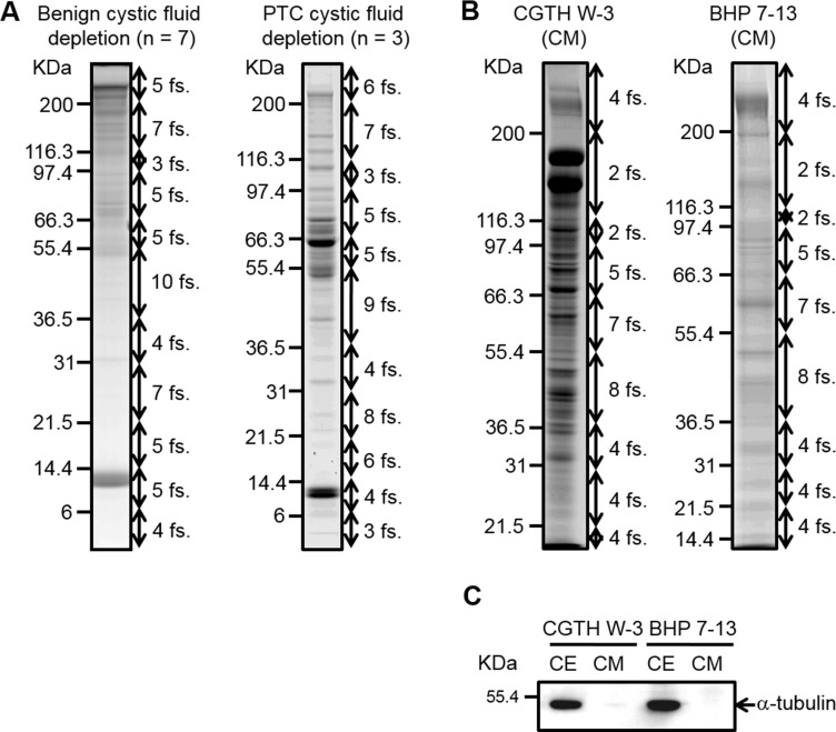
Figure 2. SDS-PAGE analysis of thyroid cystic fluid samples and conditioned media harvested from PTC cells.
(A) Thyroid cystic fluid samples of seven benign cases and three PTC cases were independently pooled and depleted using Hu-14 columns. The depleted proteins (60 μg) were resolved on 8–14%-gradient SDS gels and stained with Coomassie Blue. The gel lanes were sliced into 60 fractions (fs.) for further analysis. (B) The conditioned media (CM) of CGTH W3 and BHP 7-13 cells were collected and processed as described in the “Experimental Procedures.” Proteins (50 μg) from the concentrated CM were resolved on 8–14%-gradient SDS gels and stained with Coomassie Blue. The gel lanes were sliced into 40 pieces for further analysis. (C) Proteins (40 μg) from the CM and extracts of PTC cell lines (CE) were resolved by SDS-PAGE and subjected to Western blot analysis using an antibody against α-tubulin.

