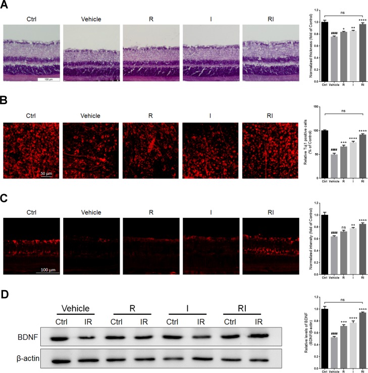
Figure 2. Retinal protection of rasagiline combined with idebenone against RIR injury.
(A) HE staining shows the thickness of retinas at day 7 after IR injury. (B) Immunofluorescent staining by Tuj1 on retinal flat mounting displays the survival of RGCs after RIR lesions. (C) Immunofluorescent staining displays BDNF levels (red) in the GCLs of the IR-injured retina. (D) Western blot analysis for the expression level of BDNF in the whole IR-injured retinas. n = 8, ####p < 0.0001, versus control; *p < 0.05, **p < 0.01, ***p < 0.001, ****p < 0.0001, versus vehicle; ns: no significance.

