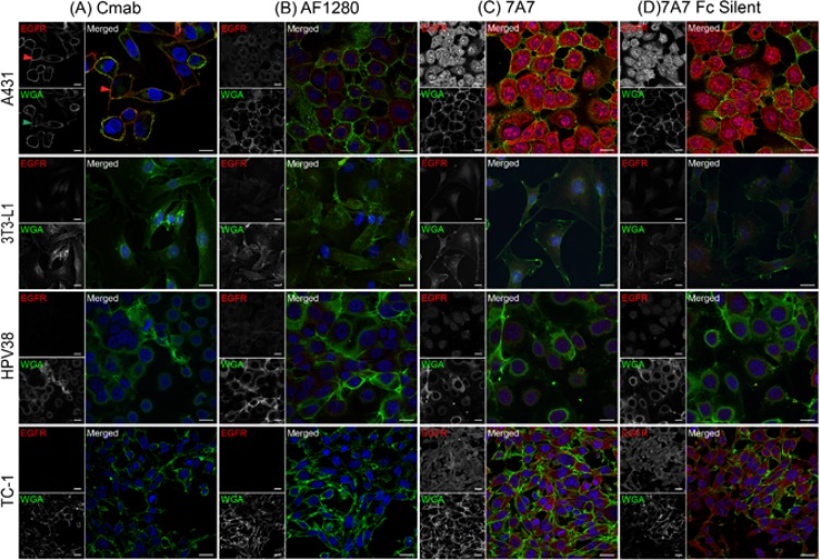
Figure 3. 7A7 and 7A7 Fc Silent do not detect EGFR expression by immunofluorescence.
Four cell lines, A431, 3T3-L1, HPV38 and TC-1 were used to assess the binding of cetuximab (A), AF1280 (B), 7A7 (C) and 7A7 Fc Silent (D) antibodies as described in Materials and Methods. Red staining: Alexa Fluor 594-conjugated secondary antibodies, Blue staining: DAPI (nucleus), Green staining: plasma membrane as indicated by wheat germ agglutinin (WGA-FITC). Cmab; Cetuximab. Scale bar is 20 µm.

