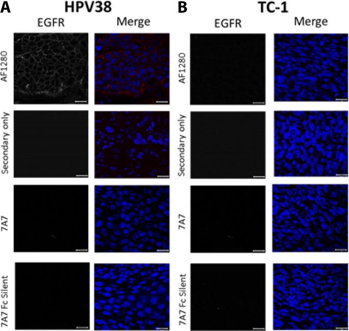
Figure 5. 7A7 and 7A7 Fc Silent do not detect EGFR expression in HPV38 tumour tissue by immunofluorescence.
EGFR expression in formalin fixed paraffin-embedded samples from HPV38 (A) and TC-1 (control; B) tumour tissues. Sections were immunostained with 3 primary antibodies (7A7, 7A7 Fc Silent and AF1280) and Alexa Fluor 594-conjugated anti-mouse or anti-goat secondary antibodies (red). Secondary only (Alexa594-conjugated goat anti-mouse IgG1 or Alexa594-conjugated donkey anti-goat IgG1) served as negative controls. Nuclei were stained using DAPI (blue). Scale bar is 20 µm.

