
Figure 3.
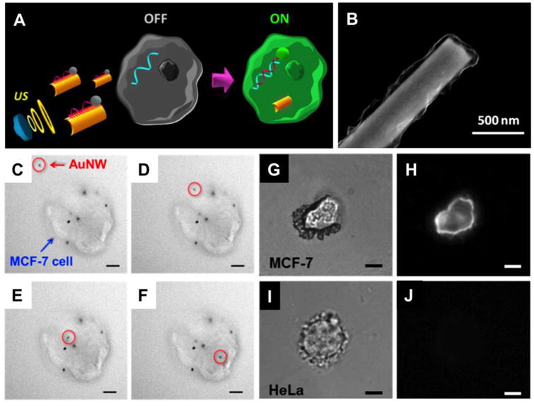
Intracellular detection of miRNAs by ultrasound-propelled ssDNA@GO-functionalized gold nanomotors. (A) Schematic illustrations of the “OFF-ON” fluorescent switching system for the specific detection of miRNA-21 in intact cancer cells. (B) Scanning electron microscopy (SEM) image of GO modified AuNW. (C-F) Actual time-lapse images at 4-s intervals illustrating the internalization process of one modified nanomotor (red circle) into a representative single MCF-7 cell (blue arrow) with some other nanomotors already internalized while some stuck on the membrane (ultrasound field, 6 V and 2.66 MHz, Scale bar, 10 μm). (G-J) Specific detection of miRNA-21 in different cell lines. Optical and fluorescence images of a single MCF-7 (G and H) and HeLa cells (I and J), respectively, after 10 min incubation with the ssDNA@GO-modified nanomotors under an ultrasound field (6 V and 2.66 MHz). Scale bar, 10 μm. (Reprinted with permission from Ref 57. Copyright 2016)