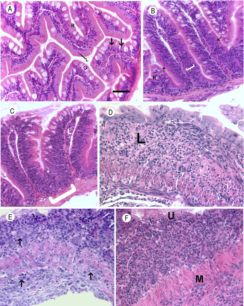
Figure 2.

Histological sections of zebrafish intestines. Hematoxylin and eosin. Bar = 25 μm. A. Normal intestine. Nuclei are basal, lymphocytes (L) are uncommon within the epithelium, and goblet cells (G) were frequently observed. B. Moderate epithelial hyperplasia in an exposed fish from group A. The layer of basal located nuclei within the mucosal epithelium is diffusely thickened (bracket) and there are numerous intraepithelial leukocytes composed primarily of lymphocytes (arrows) C. Severe epithelial hyperplasia from an exposed fish from group A. The layer of basilar nuclei is severely thickened and extends to near the brush border in some places (arrow). At the bases of intestinal folds, the mucosal epithelium is markedly thickened and bulges into the muscularis (bracket). D. Chronic severe lymphoplasmacytic and fibrosing enteritis in an exposed fish from group D. The lamina propria (L) is severely thickened by loosely organized fibrous connective tissue containing an inflammatory infiltrate Intestinal folds have been blunted or effaced, and the luminal aspect is lined by a layer of severely dysplastic epithelial cells. E. Small cell intestinal carcinoma from in an exposed fish from group B. Expanding the lamina propria is a poorly-demarcated, unencapsulated neoplasm composed of nests and clusters of polygonal cells (arrow) embedded in a scant fibrovascular stroma. Neoplastic cells have variably distinct cell borders with large amounts of infrequently granular, lightly basophilic cytoplasm. The neoplastic cells invade the tunica muscularis (M) and are present on the serosal surface of the intestine(S). F. Small cell carcinoma in an exposed fish from group B. The lamina propria contains large numbers of neoplastic epithelial cells similar to those described in image E. The overlying epithelium is ulcerated (U). Neoplastic cells invade through the tunica muscularis (M).
