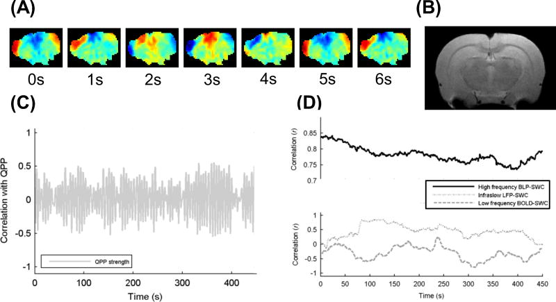
Figure 1.
Examples of different types of dynamic observed in resting state fMRI. Results are shown in coronal slices from a dexmedetomidine-anesthetized rat. (A) Filmstrip of a quasi-periodic pattern (QPP), a type of large-scale wave seen reproducibly during the resting state. (B) Anatomical image for reference. (C) Relative strength of the QPP over time. (D) Correlation between two regions of interest either for electrophysiology (black line and light gray line) or BOLD (dark gray line) in a 50 s sliding window, slid in 1 s increments across a 500 s time course. (Reprinted with permission from (Thompson et al., 2015))

