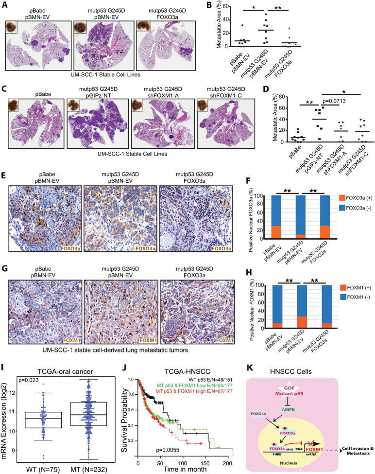
Fig. 7.
FOXO3a inhibits but FOXM1 promotes GOF mutant p53-mediated pulmonary metastasis of UM-SCC-1 cells in vivo. a, c Representative images of H&E-stained sections of lungs 13 a and 14 c weeks, respectively, after tail-vein injection of UM-SCC-1 stable cell lines. b, d Quantitation of the lung metastatic areas in a and c, respectively. *<0.05; **p < 0.01. e, g Representative images of FOXO3a e and FOXM1 g IHC staining of pulmonary metastatic lesions derived from the tail-vein injected UM-SCC-1 stable cell lines. f, h Quantitation of the positive nuclear staining of FOXO3 and FOXM1 in e and g, respectively. **p < 0.01. i FOXM1 mRNA expression of oral cancers (n = 307) with wild-type (WT) or mutant (MT) TP53 in the HNSCC TCGA dataset. 307 oral cancer samples were identified in 505 HNSCC TCGA tumor samples as tumors originating from subsites including oral cavity, oral tongue, floor of mouth, alveolar ridge, buccal mucosa, hard palate, and lip. j Survival of HNSCC patients with wild-type TP53 (WT) (n = 151), mutant TP53 (MT) and high levels or low levels of FOXM1 expression (n = 354). High or low FOXM1 expression was defined as the levels of the expression that are greater than (high) or less than (low) the median/mean expression in mutant p53 group. k Diagram of the working model of mutant p53 GOF in HNSCCs, in which GOF mutant p53s promote cell invasion and metastasis through regulation of the AMPK-FOXO3a-FOXM1 signaling axis

