Figure 3.
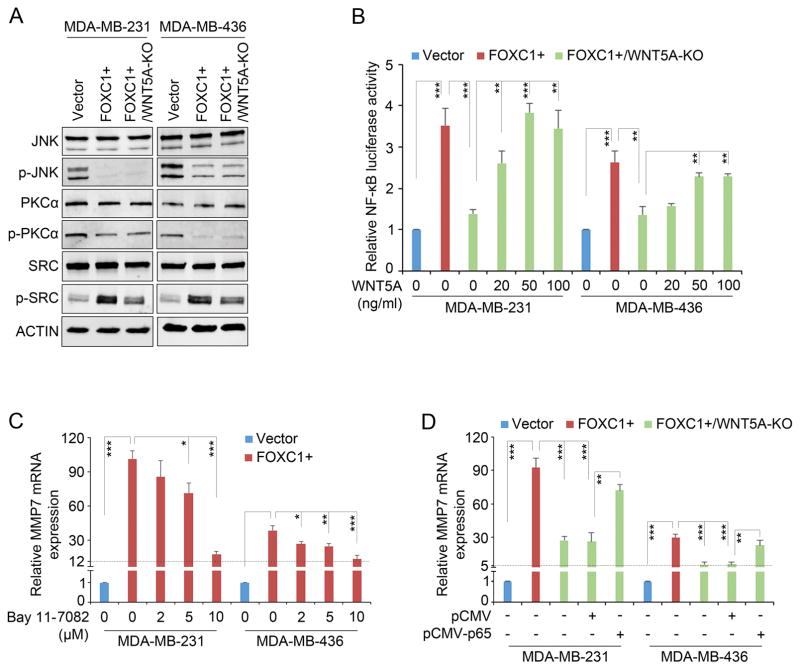
WNT5A activates NF-κB signaling in TNBC cells. A, western blotting analysis of reported WNT5A pathways in TNBC cells. ACTIN was used as an internal control. The antibodies used were from Cell Signaling Technology at 1:1000 dilution: JNK (#9252), p-JNK (#9255), PKCα (#2056), p-PKCα (#9375), SRC (#2109), and p-SRC (#6943). B, luciferase assays in the cells transfected with NF-κB-responsive luciferase reporter construct and treated with recombinant WNT5A protein at different concentrations. NF-κB responsive luciferase reporter construct pGL4.32[luc2P/NF-κB-RE/Hygro] was from Promega. The bar graph indicates mean ± SD, n = 3. **, p < 0.01, ***, p < 0.001. C and D, real-time PCR analysis of MMP7 mRNA in cells treated with the NF-κB inhibitor Bay 11-7082 (Cayman Chemical) at different concentrations (C) or transfected with the empty vector pCMV or pCMV-p65 (D). The bar graph indicates mean ± SD, n = 3. *, p < 0.05, **, p < 0.01, ***, p < 0.001.

