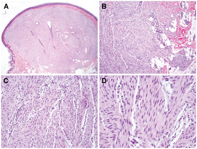
Figure 1. Histologic features of the index case, a heel tumor in a one year-old boy.
The tumor presented as a buldging nodule involving the dermis and subcutaneous tissues with infiltrative border (A,B). It is composed of intersecting fascicles of uniform plump spindle cells with fibrillary cytoplasm and bland fusiform nuclei (C,D).

