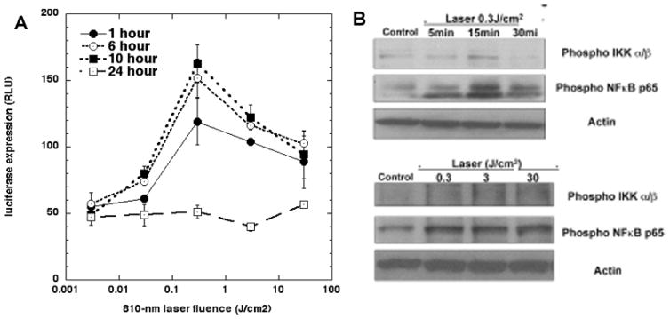
Figure 3. Activation of NF-kB (nuclear factor kappaB) in mouse embryonic fibroblasts.
Cells were isolated from NF-kB luciferase reporter mice. (A) Biphasic dose response of NF-kB activation (0.003 to 30 J/cm2 of 810 nm laser) measured by bioluminescence signal production at 1, 6, 10 and 24 hours post-PBM. (B) Western blot showing phosphorylation of NF-kB with different doses and times. Adapted from data contained in (45).

