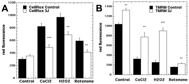
Figure 5. Effects of PBM on cells under oxidative stress.
Primary cortical neurons were treated with one of three different agents (cobalt chloride, hydrogen peroxide, rotenone) each of which produced oxidative stress. They were treated either with no PBM or with 3 J/cm2 of 810 nm laser. (A) Intracellular ROS (measured by CellRox red fluorescent probe) were modestly increased in control cells, but significantly reduced in all three types of oxidative stress. (B) In every case the mitochondrial membrane potential (measured by tetramethyl-rhodamine methyl ester fluorescent probe, TMRM) was significantly increased. Adapted from data contained in (54).

