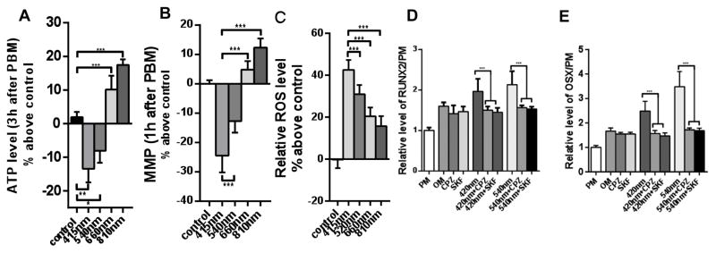
Figure 7. Effect of PBM with four different wavelengths on human adipose-derived stem cells (hADSC).
hADSCs in prolioferation medium (PM) were exposed to 3 J/cm2 of 415, 540, 660, or 810 nm light. (A) ATP measured 3 h post PBM for luciferase assay. (B) MMP measured by TMRM 1 h post PBM. (C) intracellular ROS measured by CM-H2DCFDA fluorescence probe 30 min post-PBM. (D) Expression of RUNX2 and (E) expression of osterix (OSX), both measured by RT-PCR after cells were cultured in osteogenic differentiation medium and received PBM as above every 2 days for 3 weeks. Both 415 nm and 540 nm gave significant increases in osteogenic markers that could be blocked by TRP ion channel inhibitors capsazepine, CPZ and SKF96365. 660 nm and 810 nm were less effective at osteogenic differentiation and ion channel blockers had no effect (data not shown). Partly adapted from data contained in (67).

