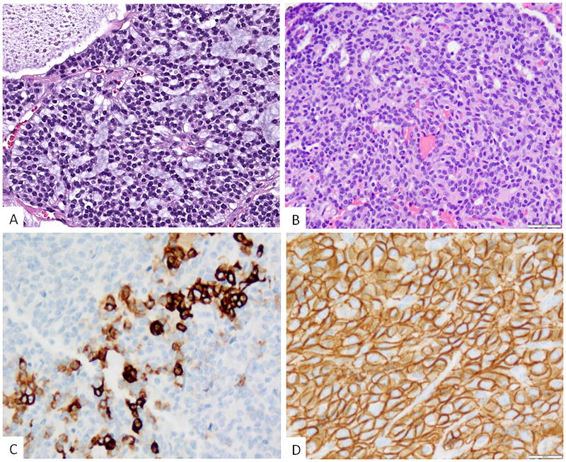
Figure 3. Morphologic features of S100 protein-negative tumors harboring ACTB1-GLI1 fusion.

A. Epithelioid neoplasm showing a distinctive cribriform or sieve-like pattern with intervening myxoid stroma (Case 5). The tumor was negative for all immunomarkers tested; B. A second tumor revealed mainly solid sheets of epithelioid cells with scattered tubular structures (case 6); immunohistochemically the lesion showed (C) focal cytokeratin staining, mainly as single cells and small clusters, most of the tumor being negative; and (D) diffuse staining for CD56.
