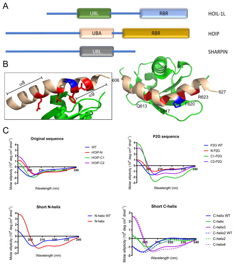
Figure 1.
(A) Illustration of LUBAC structural domains. The HOIP-UBA/HOIL-UBL interaction is indicated. (B) Crystal structure of the HOIP-UBA/HOIL-UBL interaction (PDB: 4DBG). UBA – beige, UBL – green. Red indicates key residues for binding, blue indicates P619. (B) Circular dichroism spectra in H2O of peptides presented in this work (50 μM peptide concentration).

