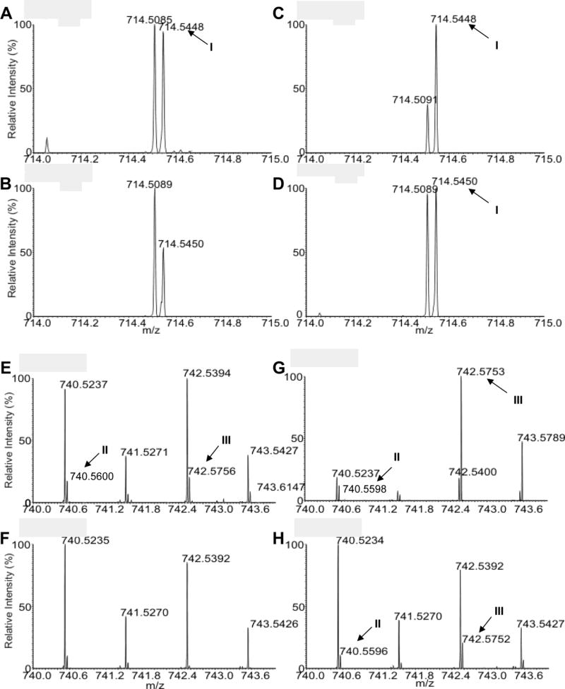
Fig. 2.
Detection of cyclopropane fatty acid (CFA)- plasmenylethanolamine (PME) in Leishmania mexicana. Total lipids from log phase promastigotes were analyzed by high resolution electron spray ionization Fourier Transform Mass Spectrometry in the negative ion mode. Mass spectra in the mass range of m/z 714.0–715.0 (A–D) and m/z 740.0–744.0 (E–H) were shown. (A, E) Leishmania mexicana wild type (WT); (B, F) cfas−; (C, G) cfas−/+CFAS; (D, H) cfas−::HA-CFAS. I-III represent the three CFA-PME species detected as [M - H]− ions, which are within 2 ppm deviation of the calculated m/z. I: p16:0/C19:0cPro(9)-PE (C40H77O7NP, calculated m/z 714.5443). II: p18:0/C19:1cPro-PE (C42H79O7NP, calculated m/z 740.5600). III: p18:0/C19:0cPro(9)-PE (C42H81O7NP, calculated m/z 742.5759). In (G), the abundance of III in cfas−/+CFAS is much higher than that of m/z 742.5400 which represents the 18:1/18:1-PE (C41H77O8NP). In contrast, the [M - H]− ions of 18:1/18:1-PE in WT (E), cfas− (F), and cfas−::HA-CFAS (H) are more abundant than III. Please note that ions at 741.5270–742.5271, 743.5426–743.5427 and 743.5789 are 13C1-isotopmers.

