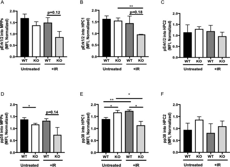
Figure 4. DEK KO progenitor cells demonstrate decreased Erk1/2 and p38 phosphorylation in response to irradiation.
(A–C) Levels of phosphorylated Erk1/2 were modestly downregulated in irradiated DEK KO (A) MPP and (B) HPC1 cells but not (C) HPC2 cells compared to DEK WT cells. (D-F) Levels of phosphorylated p38. (D) In MPP cells were significantly decreased in DEK KO (D) MPP cells compared to WT mice. (E) In the HPC1 subpopulation, untreated DEK KO cells upregulated phosphorylation of p38, which was aberrantly downregulated following irradiation. (F) HPC2 cells did not demonstrate differences in phosphorylated p38 levels between DEK WT and KO cells in either condition. The graphs depicts mean fluorescence intensity (MFI) as determined by flow cytometry. N=4 for each group of untreated animals and N=3 for each group of irradiated animals.

