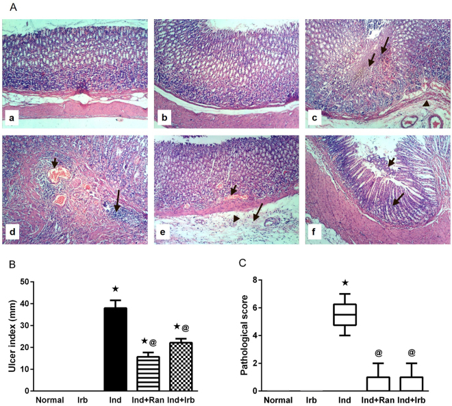
Figure 2.
Effect of irbesartan on gastric mucosal damage induced by indomethacin in rats. (A) Histological assessment of gastric tissues using H&E stain (X 100). (a) Normal group showed normal histology of gastric layers. (b) Irb group showed no histopathological changes in the gastric layers. (c) Ind group showed focal coagulative necrosis of gastric mucosa (small arrow); associated with inflammatory cells infiltration (large arrow) and submucosal edema (arrow head). (d) Ind group showed congestion of submucosal blood vessels (small arrow) and massive submucosal inflammatory cells infiltration (large arrow). (e) Ind + Ran group showed mild signs of congestion of mucosal blood vessel (small arrow) and submucosal edema (large arrow) as well as few submucosal inflammatory cells infiltration (arrow head). (f) Ind + Irb group showed mild focal necrosis and sloughing of gastric mucosa (small arrow) beside infiltration of few inflammatory cells (large arrow). (B) Ulcer index. Each bar with vertical line represents the mean ± S.E.M of 6 rats. *vs normal, @vs Ind (one-way ANOVA followed by Tukey’s multiple comparisons test; p < 0.05). (C) Pathological score. Each bar with vertical line represents the median ± range of 6 rats. *vs normal, @vs Ind (Non-parametric Kruskal-Wallis one-way ANOVA followed by Dunn’s multiple comparisons test). Irb, Irbesartan; Ind, indomethacin, Ran, ranitidine.

