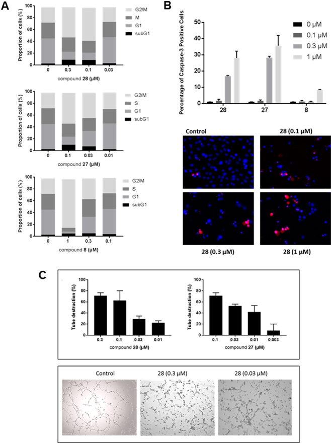
Figure 6.
Mechanism of action of the benzofurane derivatives. (A) Inhibition of cell cycle progression. MDA-MB-231 cells were treated with DMSO (control) or compounds 8, 27 or 28 for 24 h. Next, the cells were harvested, stained with propidium iodide (PI), and cell cycle distribution was evaluated by flow cytometry. Percentages of cells in the different phases of the cell cycle are indicated. (B) Caspase-3 activity. MDA-MB-231 cells were seeded in 48-well plates at 40,000 cells/cm². After 24 h, different concentrations of compounds 8, 27 or 28 and 2 µM of the caspase-3 substrate DEVD-NucView488 were added. After 16 h, the cells were incubated with 2 µg/ml Hoechst 33342 to stain the nucleus, and imaged. (C) Disruption of the vascular network. HMEC-1 cells were cultured on matrigel for 3 h to allow the formation of tube-like structures. Next, different concentrations of compounds were added. Upper panel: After 90 min, pictures were taken and the tubular network was scored (3: intact network, 0: network completely destroyed). Bars show average ± SEM (n = 4). Lower panel: Microscopic pictures of the tubular network 3 h after addition of compound.

