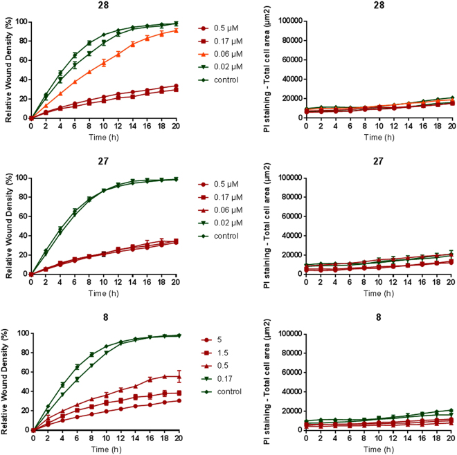
Figure 7.
Inhibition of HMEC-1 migration. The 96-well IncuCyte® scratch wound assay was used to create a cell-free zone (wound) in a confluent cell monolayer. Next, compounds were added and the relative wound density was measured every minute and visualized in time-course plots (left panels). PI is added to all wells to visualize toxicity over time, i.e. only dead cells take up this cell-impermeable dye (right panels). Average ± SEM (n = 3) are shown.

