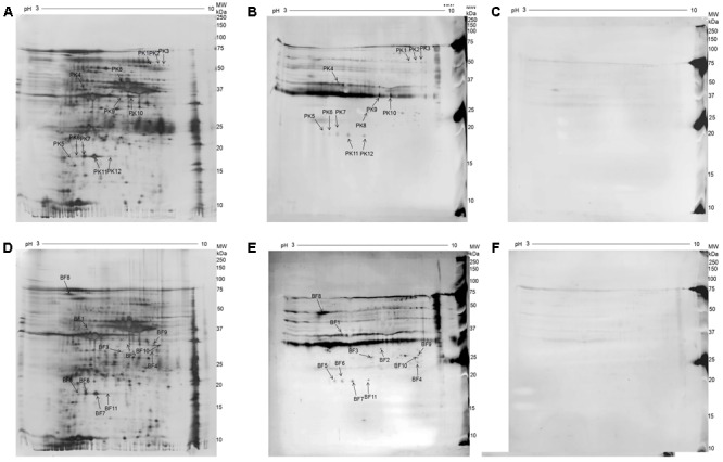
FIGURE 2.

2D electrophoresis (2-DE) gel and Western blot analysis of whole-cell proteins of M. bovis 08M grown as planktonic cells and biofilms. (A) Silver-stained 2-DE gel of whole-cell proteins from M. bovis grown as planktonic cells (pH 3–10, 13 cm). (B) Western blot analysis of whole-cell proteins from M. bovis grown as planktonic cells using convalescent sera against M. bovis 08M. (C) Western blot analysis of whole-cell proteins from M. bovis grown as planktonic cells using pre-infected sera. (D) Silver-stained 2-DE gel analysis of whole-cell proteins from M. bovis grown as biofilms (pH 3–10, 13 cm). (E) Western blot analysis of whole-cell proteins from M. bovis grown as biofilm using convalescent sera against M. bovis 08M. (F) Western blot analysis of whole-cell proteins from M. bovis grown as biofilm using pre-infected sera.
