Fig. 1.
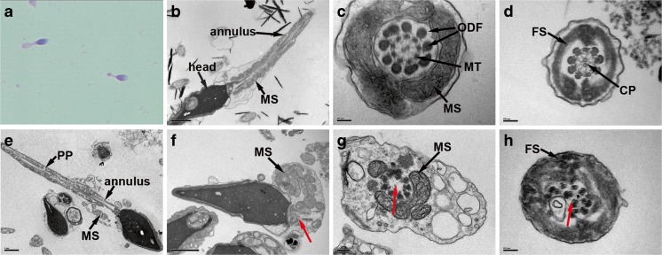
Morphological observation on sperm flagella. a Light microscopy observation of the man affected with multiple morphological abnormalities of the sperm flagella (MMAF) shows short and thick sperm flagella. Transmission electron microscopy (TEM) observation on morphologically normal spermatozoa (b–d). b A longitudinal section showed regular arranged flagellar mitochondrial sheath (MS) in the middle piece and normally formed annulus. c A cross section at middle piece displayed a normal axoneme with a 9 + 2 arrangement of nine peripheral microtubules (MT) and central pair of microtubules (CP), surrounded by nine outer dense fibers (ODF) and MS. d A cross section at principal piece (PP) showed a well-organized axoneme surrounded by normal fibrous sheath (FS). TEM observation on the MMAF affected man demonstrated several ultrastructure anomalies in the flagella (e–h). e The MS was badly assembled, and the annulus was abnormally located. In addition, the FS in principal piece was thickened and unsmooth. f The MS together with all other flagellar structures was unassembled, but the proximal centriole and the segmented columns were normally formed (red arrow). g The CP was absent at middle piece (red arrow), and the MS was incomplete. h The section at principal piece showed disorganized microtubules (red arrow) and thickened FS. Scale bars = 1 μm (b, e, f), 0.1 μm (c, d, h), and 0.2 μm (g)

