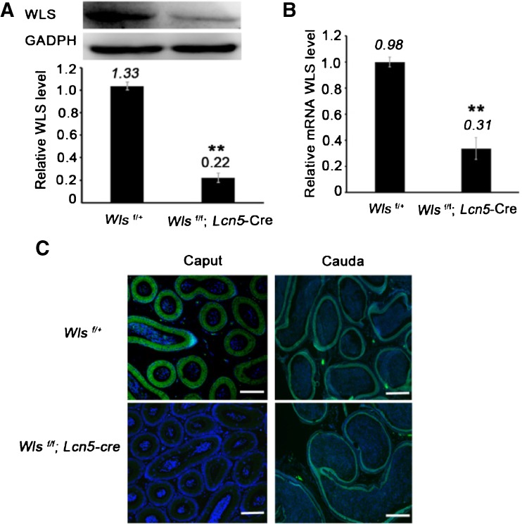
Fig. 2.
Wls deletion in caput epididymidis. a Detection of WLS in caput epididymidis using Western blot analysis. The protein level was normalized to GAPDH. b Wls mRNA abundance was detected using qPCR and was normalized to Gapdh. c Immunostaining of WLS in caput and cauda epididymides. Bar = 100 μm. DNA, blue; WLS, green. In a, b, ** P < 0.01; the presented data are from ten control or ten conditional knockout males; the data are shown as the mean ± standard error. In a–c, caput epididymides were collected from middle/distal caput epididymides of 2-month-old Wls f/+and Wls f/f ; Lcn5-Cre mice

