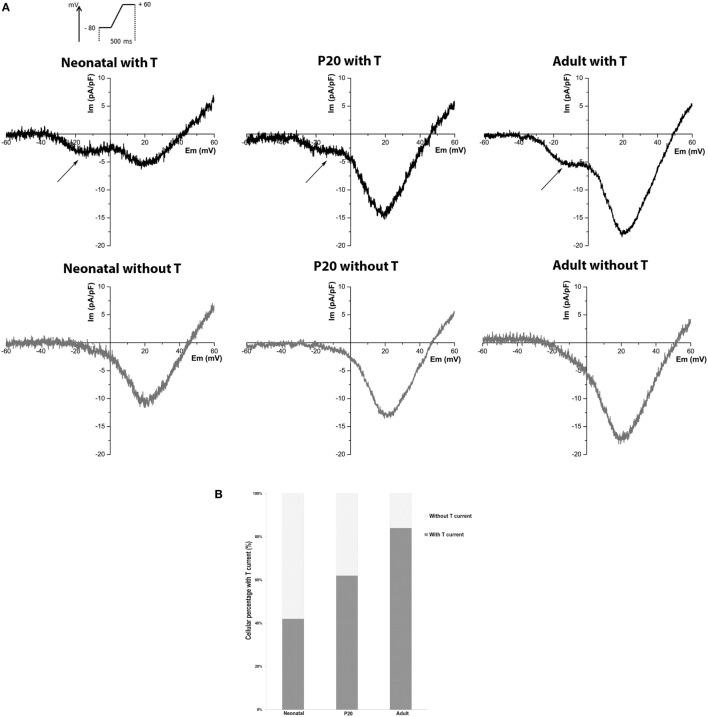
Figure 1.
Detection of T-type current in neonatal, P20, and adult beta cells. (A) Representative recordings of global calcium currents observed in neonatal, P20, and adult beta cells. Arrows represent T-type calcium current. (B) Quantification of the beta-cell percentage with and without T-type calcium current in neonatal (n = 76), P20 (n = 78), and adult (n = 86) beta cells.

